What is Arthroscopic Surgery?
In arthroscopic surgery, a small camera is inserted into the joint through a tiny incision, and the image is transmitted to a monitor. The surgeon then performs the operation by watching the monitor and using various surgical instruments inserted through another small incision into the joint. The surgical instruments used in arthroscopic surgery are as small as a pen tip.
What Are Its Advantages?
- The meniscus, cartilage, and ligaments inside the joint are tissues that do not have pain receptors. Arthroscopic surgery allows direct entry into the joint without damaging or injuring the surrounding tissues, enabling intervention only on the affected tissues. As a result, the patient experiences minimal discomfort after the operation.
- During arthroscopic surgery, the internal structures of the joint are viewed up close and magnified, allowing for a much better diagnosis and treatment of diseased tissues.
- In arthroscopic surgery, the healing process is facilitated as the muscles and capsule that enable joint movements, which are usually difficult and painful to heal, remain untouched. Consequently, postoperative joint movements are painless and comfortable, and the risk of infection is significantly lower compared to open surgeries.
What Are the Applications of Knee Arthroscopy?
Arthroscopic surgery can be used to treat almost all joint-related conditions. The most common applications include:
- Meniscus surgery
- Anterior and posterior cruciate ligament operations
- Some cartilage diseases of the joint
- Early-stage knee osteoarthritis
- Patellar misalignment and dislocations
- Intra-articular infections
- Benign tumors and cysts inside the knee joint
Arthroscopic Meniscus Surgery
Meniscus injuries are the most common indications for knee arthroscopy. The medial meniscus is more frequently torn than the lateral meniscus. Meniscus injuries are rare in childhood. In individuals over 50 years of age, meniscus tears typically occur due to degeneration rather than trauma.
Clinically, patients with meniscus injuries may experience joint pain, swelling, popping sounds in the knee, locking, and instability. Meniscus tears can occur alone or in combination with other knee injuries (e.g., anterior cruciate ligament injuries). Some meniscus tears may become trapped between bones, preventing full extension of the knee.
For small, stable meniscus tears that do not cause clinical symptoms, surgical treatment is not necessary. However, in cases where symptoms persist, arthroscopic intervention is required. The goal of arthroscopy is to remove only the torn part of the meniscus or, if possible, to repair it. There are two types of surgical approaches for meniscus treatment. If the tear is in a region without blood vessels, only the torn section is removed. If the tear is in a vascularized area (the "red zone") and the patient is under 45 years of age, the tear is repaired with sutures. However, in cases where red-zone tears are old, fragmented, or occur in individuals over 45, suturing is not preferred, and the torn section is removed instead.
Meniscus repair is a highly complex and challenging surgery that should be performed only by experienced knee surgeons. In some cases, an additional small incision may be necessary to protect the nerves and blood vessels at the back of the knee during lateral meniscus repair.
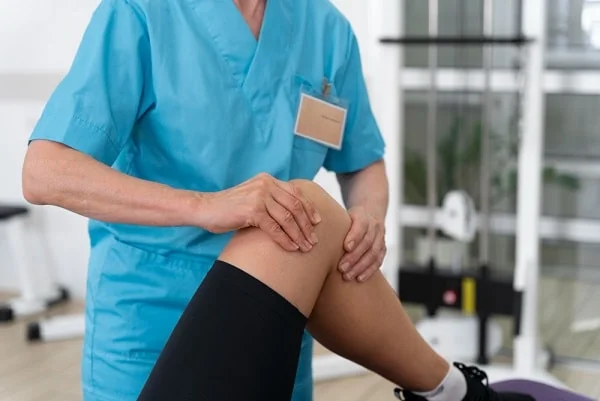
After meniscus surgery, patients are usually discharged the next day. There is minimal postoperative pain. If the meniscus tear is removed, patients can walk without crutches and go up and down stairs immediately after surgery. Two days of ice application and home rest are recommended, after which patients can return to office work. Those with physically demanding jobs can return to work on the 10th day. Light running can begin on the 20th day, and full sports activities can be resumed at the end of the month. Professional athletes may return to sports as early as the 20th day with specialized rehabilitation programs.
What Are Arthroscopic Cartilage Operations?
Arthroscopic surgery offers several treatment options for cartilage damage in the knee joint. Below are summarized procedures:
- Microfracture Technique: The bone marrow contains cells capable of transforming into cartilage cells. In this method, small fractures are created in the damaged cartilage area to stimulate the formation of new cartilage tissue. The newly formed cartilage differs slightly from the original cartilage. Postoperatively, patients can bend their knees comfortably but must avoid putting weight on the leg for 6-8 weeks, using crutches instead. An intensive physical therapy program is required for the following six weeks.
- Shaving Technique: A commonly used method that involves shaving and smoothing irregular cartilage surfaces. This technique does not promote new cartilage formation but reduces friction, wear, and pain by improving surface smoothness. It is a simple procedure that requires delicate execution. Postoperative recommendations are similar to those for meniscus surgery.
- Osteochondral Grafting (Mosaicplasty): This procedure involves transplanting cylindrical cartilage and bone grafts from healthy joint areas to damaged regions. Autografts (the patient’s own tissue) are preferred whenever possible. The grafts are taken from low-weight-bearing joint surfaces, which limits the size of the treated area. These grafts are placed in a mosaic pattern in the damaged area. This method is effective for weight-bearing areas with relatively small defects and provides a near-original durable joint surface. Postoperatively, early movement and strengthening exercises are encouraged, but weight-bearing is restricted for 6-8 weeks. Full weight-bearing is delayed up to three months, and return to sports takes 4-6 months.
- Cartilage Cell Transplantation: Cartilage cells are highly differentiated and cannot multiply on their own. In humans, cartilage cell production ceases at around one year of age. To generate new cartilage cells, genetic procedures and cell culture techniques are required. This procedure is performed in two stages. In the first stage, the surgeon collects healthy cartilage cells arthroscopically from non-weight-bearing areas of the knee joint. These cells are then cultured in a laboratory for about 15 days. Once sufficient cells are produced, the second stage of surgery is performed, during which the cultured cells are implanted under a stitched periosteal cover in the damaged cartilage area. The resulting cartilage tissue closely resembles original cartilage. Since the patient’s own cells are used, there is no risk of rejection, and approximately 70-80% of patients achieve significant improvement. However, this procedure is not suitable for all patients. Factors such as the size of the damaged area, previous surgeries, patient expectations, lesion location, and multiple lesions must be considered. It is not recommended for elderly patients or those with generalized osteoarthritis but is suitable for young patients with post-traumatic cartilage defects.
The postoperative protocol includes early movement, strengthening exercises, and avoiding weight-bearing for 6-8 weeks. Full weight-bearing is delayed up to three months, while return to sports activities takes 6-8 months.
Op. Dr. Yılmaz Şahin
Orthopedics and Traumatology Specialist