
After completing my six-year medical education at Ankara University Faculty of Medicine, I completed my Radiology residency at the Radiology Department of Ondokuz Mayıs University (OMÜ).
I have been a Radiology specialist for 11 years. For the last 9 years of my specialty career, I worked at Samsun Maternity and Children’s Hospital. Throughout my professional experience, I have observed that in many diseases, the path to diagnosis passes through radiology.
In Radiology, the primary and, in some cases, the only diagnostic method used is ultrasonography. Although ultrasound has traditionally been described as “easily accessible,” in today’s world, patients face increasing difficulties in accessing ultrasound services.
With the experience I have gained over the years, I established my private practice to provide a solution to this issue, offering high-quality healthcare services under optimal conditions. Using our state-of-the-art, next-generation ultrasound device, we conduct patient examinations, and reports are immediately delivered as a file upon completion of the procedure.
What is Ultrasonography?
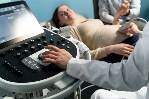
Ultrasonography is a medical imaging technique that uses high-frequency sound waves from an ultrasound device to visualize the body. Since it does not contain X-rays, which are known as radiation, it is a safe, reliable, and easily accessible method. It is especially the first-choice diagnostic tool for pregnancy monitoring, as well as for infants and pediatric patients.
How is Ultrasonography Performed? How Long Does It Take?
During an ultrasound procedure, a water-based substance known as ultrasound gel is applied to the area to be examined. A probe is then moved over the area, and the images are monitored on a screen. The duration of the procedure varies depending on the organ or region being examined, typically ranging from 10 to 60 minutes.
Which Organs Can Be Evaluated with Ultrasonography?
- Abdominal region: Liver, gallbladder, spleen, pancreas, kidneys, urinary bladder, intestines, uterus, and ovaries in women, prostate in men
- Breast
- Neck region: Thyroid gland, salivary glands, lymph nodes
- Testicles and scrotum in men
- In infants: Hip ultrasound for hip dysplasia and transfontanel ultrasound for brain evaluation
- Follicular monitoring in patients planning pregnancy using transvaginal ultrasound
- Color Doppler Ultrasonography (CDUS) evaluates blood flow in the vessels. It is most commonly used to assess conditions such as vascular occlusions and varicose veins in the legs, vascular blockages in the arms, hemodialysis fistulas, and narrowing or blockages in the neck vessels. Additionally, it is utilized to evaluate blood flow in all organs and masses.
How Often Can Ultrasonography Be Performed?
Ultrasonography can be performed as often as your doctor deems necessary. In cases requiring close monitoring, the same patient can undergo multiple ultrasound examinations within a single day. There are no known proven risks associated with ultrasound to date.
What is a Detailed Ultrasound in Pregnancy? When Is It Performed?
One of the most common and effective uses of ultrasonography is Obstetric Ultrasound, also known as Pregnancy Ultrasound. The most crucial ultrasound examinations that must be performed on every pregnant woman include:
- First Trimester Detailed Ultrasound Screening (between 11-14 weeks)
- Second Trimester Detailed Ultrasound Screening (between 20-24 weeks)
Beyond these, ultrasound follow-ups continue at varying intervals until birth.
With detailed ultrasound, the baby’s brain, face, chest cavity, heart, internal abdominal organs, spine, hands, arms, feet, legs, gender, blood vessels supplying the baby, placenta, amniotic fluid, uterine walls, and cervix are thoroughly examined for any abnormalities.
4D Color Ultrasonography
4D Color Ultrasonography is not the same as detailed ultrasound.
Detailed ultrasound is primarily performed using “2D Ultrasound”, which provides grayscale images.
4D Ultrasound, on the other hand, allows us to observe the baby’s external appearance, displaying the baby’s face, hands, feet, body, and position in the womb in a skin-tone-like color. It also enables recording the baby’s movements as a video.
Specialist Dr. Tuba BAYRAK ULUIŞIK
Radiology Specialist
